Eye Care in Sealy, TX: When Standard Lenses Are Not Enough
Eye care in Sealy, TX can involve more than updating a glasses prescription when vision remains distorted or contact lenses are difficult to wear. For…

Complete eye health evaluations create more value when each examination becomes part of a continuous record rather than an isolated check of visual clarity.
An eye exam produces several kinds of information. Some measurements describe how clearly a person sees. Others document the condition of the cornea, lens, retina, optic nerve, eyelids, and surrounding tissues. Medical history, symptoms, medications, and family risk add essential context.
The greatest insight often comes from comparing these findings over time. A result that appears normal today can establish a baseline for tomorrow, while a subtle change may become more meaningful when an optometrist can review earlier measurements or retinal images.
Visual acuity is important, but it is only one part of the system. Reading a 20/20 line does not rule out every eye condition, just as a change in prescription does not automatically indicate disease.
A thorough evaluation generally considers three connected layers:
These layers prevent individual results from being interpreted in isolation. Eye pressure, for example, is useful when assessing glaucoma risk, but it is not a complete glaucoma test by itself. Retinal photography may document the back of the eye, but an image still requires clinical interpretation and may not replace dilation or specialized testing.
The Centers for Disease Control and Prevention notes that regular eye care can help detect problems early, including conditions that may progress before noticeable vision loss develops.
In business systems, a single report provides a snapshot. A series of consistent reports reveals a trend. Eye care follows the same basic principle.
Baseline information may include:
At a later exam, the optometrist can evaluate whether a finding appears stable, newly developed, or gradually changing. This does not mean every variation signals a problem. Measurements can be influenced by testing conditions, device differences, fatigue, technique, and normal biological variation.
The clinician’s role is to determine whether a change is repeatable, clinically meaningful, and consistent with other evidence. Technology contributes data, while professional judgment turns that data into a care decision.
The retina is the light-sensitive tissue at the back of the eye. It contains the macula, blood vessels, and the visible portion of the optic nerve. Retinal imaging can create a permanent visual record of these structures.
A photograph may help document features such as:
Images are particularly useful for comparison. If a feature is present in both the current and prior photograph without meaningful change, that stability may provide useful context. If something appears different, the optometrist can decide whether repeat imaging, dilation, optical coherence tomography, visual field testing, or referral is appropriate.
Retinal photography and dilation serve related but different purposes. The National Eye Institute’s dilated eye exam guidance explains that widening the pupil helps the doctor examine more of the retina and other internal structures. The testing plan should be based on individual needs rather than assuming one technology replaces every other method.
Many people seek an eye exam because text has become blurry or an existing prescription no longer feels comfortable. Those are valid reasons, but ocular disease does not always announce itself through obvious blur.
Glaucoma can damage the optic nerve and often progresses without early symptoms. Peripheral vision may be affected gradually, making the change difficult to notice without testing.
The National Eye Institute explains that glaucoma evaluation may involve a dilated examination and visual field testing. Eye pressure is relevant, but people can develop glaucoma at different pressure levels. This is why optic nerve appearance, family history, age, visual fields, and other findings must be considered together.
Diabetes can damage small blood vessels in the retina. Early diabetic retinopathy may not cause symptoms, even while retinal changes are developing.
The National Eye Institute’s diabetic retinopathy resource recommends comprehensive dilated eye examinations for people with diabetes. Blood sugar control, blood pressure, cholesterol, duration of diabetes, and pregnancy can all influence risk and monitoring needs.
Age-related macular degeneration affects the macula, the retinal area responsible for detailed central vision. Early stages may not create noticeable symptoms.
According to the National Eye Institute, risk increases with age and is also influenced by factors such as smoking and family history. New distortion, including straight lines appearing wavy, deserves prompt evaluation.
A cataract is a cloudy area in the eye’s natural lens. Early changes may be subtle, while later symptoms can include faded color, glare, halos, blurred vision, and difficulty seeing at night.
The National Eye Institute’s cataract overview explains that cataracts are common with age but may also be associated with diabetes, eye injury, steroid use, smoking, and other factors. Monitoring helps connect structural changes with their effect on everyday activities.
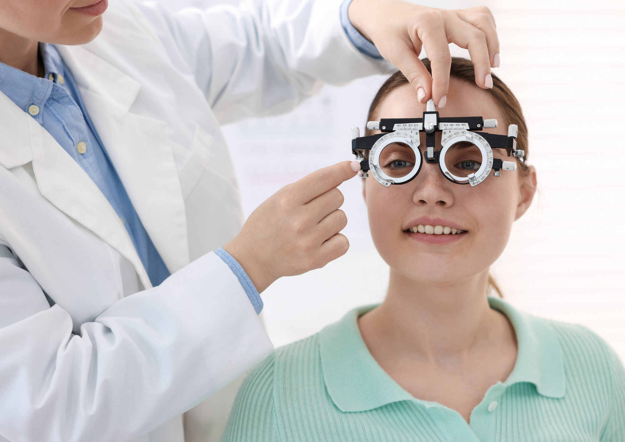
Refraction determines the lens power that helps light focus more accurately on the retina. It is the familiar part of an exam in which the patient compares different lens choices.
The result should account for more than the smallest letters that can be read. The optometrist may also consider:
A stronger prescription is not automatically a better prescription. The most useful correction is one that provides clear, comfortable vision for the person’s actual tasks.
The American Optometric Association’s eye exam information distinguishes comprehensive care from limited screenings. A screening can identify that someone may have difficulty seeing, but it does not provide the same evaluation of prescription needs and ocular health.
Not every patient needs every diagnostic test at every visit. A well-designed examination uses the history and initial findings to determine where additional attention is warranted.
Factors that may affect testing frequency or scope include:
This is similar to a risk-based monitoring system. Routine data establishes the normal operating range, while symptoms or abnormal findings trigger more targeted evaluation. The model avoids both extremes: performing every possible test without a reason and relying on a basic vision check when more information is needed.
Advanced instruments can measure and document the eye with impressive precision. However, the device does not know that a patient recently began a new medication, has a parent with glaucoma, experiences headaches after two hours of screen work, or noticed a sudden increase in floaters.
That context changes how data is interpreted. Before the exam, patients should be prepared to discuss:
A symptom timeline is especially useful. Note whether the issue began suddenly or gradually, affects one eye or both, remains constant or intermittent, and occurs during particular activities.
Nova Optique & Eyecare states that its annual examination includes an ocular health assessment, evaluation of the lashes and lids, an eyeglass prescription, and retinal images. Its published information also notes that the optometrist checks for concerns such as glaucoma, macular degeneration, and retinal holes or tears.
The practice advises that a comprehensive exam may take approximately 30 minutes to one hour, depending on the patient’s needs. That range reflects an important reality: an efficient exam is not necessarily an identical exam. Time and testing should respond to the information being gathered.
Sudden vision loss, a curtain-like shadow, new flashes accompanied by many floaters, significant eye pain, chemical exposure, or an eye injury should not wait for a routine examination. These symptoms may require urgent evaluation.
Likewise, persistent distortion, double vision, light sensitivity, or unexplained redness deserves professional guidance even if visual acuity still seems acceptable.
A prescription may be the most visible outcome of an eye exam, but the deeper value is an organized record of visual function, ocular structures, risk factors, and change over time.
Technology improves this process by creating precise measurements and comparable images. Clinical expertise adds context, recognizes patterns, and determines what deserves further attention. When both elements are used together, an eye exam becomes more than a yearly task. It becomes a practical information system for protecting vision over the long term.