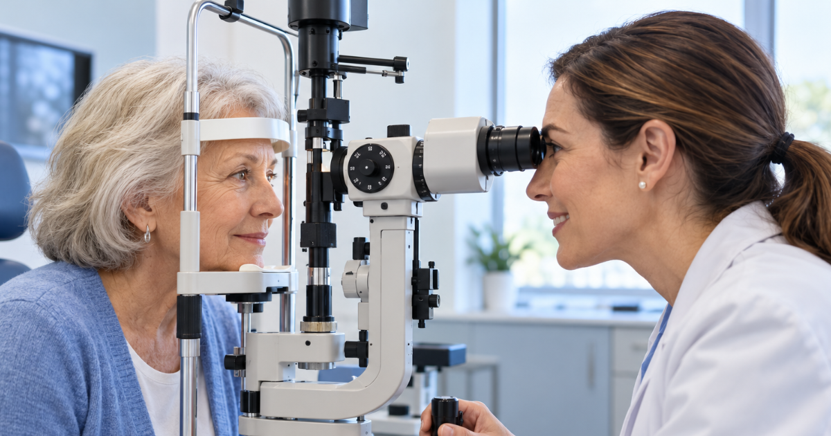
Eye Care in Sealy, TX: When Standard Lenses Are Not Enough
Eye care in Sealy, TX can involve more than updating a glasses prescription when vision remains distorted or contact lenses are difficult to wear. For…

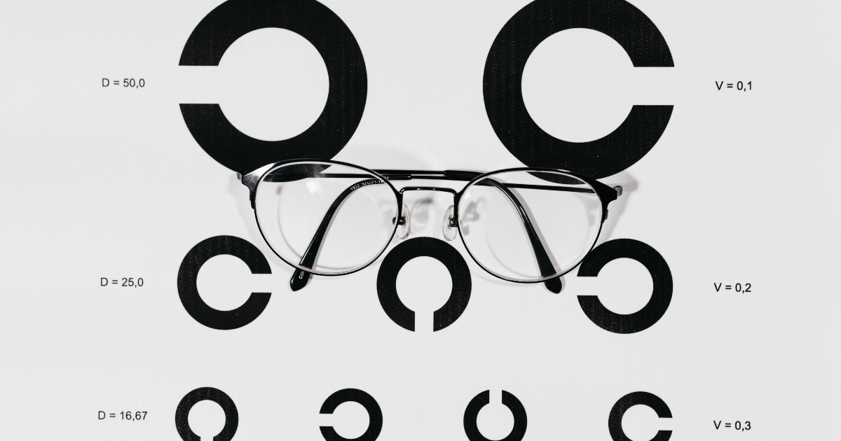
Macular degeneration care in Chino, CA is increasingly supported by imaging technology that helps eye doctors examine the retina, document subtle findings, and monitor changes that may not be immediately noticeable to patients.
This matters because age-related macular degeneration, commonly called AMD, can begin without obvious symptoms. As the condition progresses, it may interfere with the central vision used for reading, driving, recognizing faces, and completing detailed tasks.
Technology cannot replace an experienced eye doctor, but it can provide valuable information during an examination. When imaging, clinical evaluation, patient education, and appropriate referrals work together, patients have a clearer path for protecting their remaining vision.
The macula is a small, specialized area near the center of the retina. Although it occupies only part of the retina, it is responsible for the sharp central vision needed to see fine details.
Macular degeneration generally affects central vision rather than peripheral vision. It does not usually cause complete blindness, but advanced changes can make many everyday activities significantly more difficult.
The National Eye Institute identifies AMD as a leading cause of vision loss among older adults. The organization also notes that some people with early AMD may not realize they have the condition because symptoms can be absent at first.
This is one reason routine comprehensive eye examinations remain important, particularly for adults with age-related or lifestyle risk factors.
Macular degeneration is commonly discussed in two forms: dry AMD and wet AMD. Understanding the distinction helps explain why monitoring and prompt evaluation are both necessary.
Dry AMD is the more common form and often progresses gradually. It is associated with changes in the macula, including the presence of deposits called drusen. Not everyone with drusen develops advanced vision loss, so the findings must be interpreted in the context of a complete eye examination.
Wet AMD is less common but can lead to faster and more serious changes in central vision. It occurs when abnormal blood vessels grow beneath the retina and leak blood or fluid.
Sudden distortion, a new central blur, or a dark area in the center of vision requires prompt attention. The American Academy of Ophthalmology provides additional information about the forms, symptoms, and management of AMD.
A traditional eye examination provides essential information, but modern imaging can give eye doctors another way to evaluate retinal health. Images may create a visual record that can be compared across future appointments.
This comparison is particularly useful for conditions that can change gradually. A patient may not notice a small difference in vision from one month to the next, while documented retinal findings can help the doctor determine whether closer monitoring or additional evaluation is appropriate.
Digital retinal imaging captures photographs of structures at the back of the eye. These images can help document the appearance of the retina, optic nerve, blood vessels, and macular area.
Retinal photographs do not replace every other component of an examination. Depending on the patient’s symptoms and findings, dilation or additional testing may still be recommended. The value of the image comes from combining it with medical history, visual testing, and the eye doctor’s clinical assessment.
Optical coherence tomography, or OCT, is another technology used in eye care. It produces detailed cross-sectional images of retinal layers using light rather than radiation. Availability and clinical use vary among practices, and the test is ordered when an eye doctor determines it is appropriate.
An overview from the National Library of Medicine describes how OCT is used to evaluate retinal anatomy and support the assessment of several eye conditions.
For AMD, detailed imaging may help qualified clinicians evaluate structural changes, identify fluid, and monitor findings over time. The results must always be interpreted by an eye-care professional.
A single image provides information from one moment. A series of images can create a timeline.
Longitudinal monitoring allows the eye doctor to compare current findings with earlier records. This can make conversations about stability or progression more specific and easier for patients to understand.
Effective tracking may include:
Technology supports this process, but consistent follow-up is what gives the information context. Patients should attend recommended appointments even when their vision seems unchanged.
AMD symptoms can vary according to the form and stage of the condition. Some changes develop slowly, while others appear more suddenly.
Possible warning signs include:
A new visual change should not be dismissed as a normal part of aging. The MedlinePlus macular degeneration resource offers a useful overview of symptoms, diagnosis, and treatment information from the National Library of Medicine.
Sudden central distortion, a new dark spot, or rapid loss of vision should be evaluated promptly.
Age is a major risk factor for AMD, but it is not the only consideration. Family history, smoking, cardiovascular health, and other personal factors may influence risk.
Smoking is one modifiable concern. The Centers for Disease Control and Prevention explains that smoking can increase the risk of age-related macular degeneration and other eye diseases.
Patients should give their eye doctor an accurate health and family history. Useful information may include:
This information helps the doctor decide how closely the eyes should be evaluated and monitored.
Some patients are advised to monitor their central vision between appointments with an Amsler grid. The grid contains straight horizontal and vertical lines with a central point for fixation.
When used correctly, it may help a patient notice new distortion, missing areas, or changes between the two eyes. It is not a diagnostic test and should not replace scheduled examinations.
The American Academy of Ophthalmology explains how home monitoring may be used by people with AMD. Patients should follow their own doctor’s instructions and report a new change promptly rather than waiting for the next routine visit.
There is no single management plan for every person with macular degeneration. Recommendations depend on whether the condition is dry or wet, its stage, examination findings, and the patient’s overall health.
Management may involve regular monitoring, lifestyle guidance, nutritional discussions, or coordination with a retina specialist. When wet AMD is suspected, timely referral is important because specialist treatment may be required.
Some patients with certain stages of AMD may be advised to consider a specific nutritional supplement formulation studied in the Age-Related Eye Disease Studies. Supplements are not appropriate for everyone and should not be started solely on the basis of online information.
The National Eye Institute provides a detailed summary of the AREDS and AREDS2 clinical trials. Patients should discuss the findings with an eye doctor who understands their diagnosis, health history, and current medications.
Advanced equipment is helpful only when patients understand what the findings mean. An eye-care visit should provide opportunities to ask questions about risk, testing, recommended follow-up, and signs that require urgent attention.
Oasis Optometry combines personalized care with modern eye-care technology for patients in Chino, California. Its macular degeneration services include comprehensive eye-health evaluation, retinal imaging, dilation when needed, ongoing monitoring, and coordination with a retina specialist when appropriate.
This combination of technology and communication can make a complicated diagnosis easier to navigate. Images can support the evaluation, while a clear discussion helps patients understand what they should do next.
When central vision has already been affected, additional support may help patients continue performing important daily activities. Vision rehabilitation can include lighting changes, magnification, adaptive devices, and strategies for using remaining vision more effectively.
The National Eye Institute’s vision rehabilitation resources provide information for people experiencing vision loss and for the family members who support them.
Seeking rehabilitation is not the same as giving up on medical care. It is a practical way to address daily challenges while continuing to follow the treatment or monitoring plan established by eye-care professionals.
The greatest advantage of modern eye-care technology is not simply that it produces detailed images. Its real value is the ability to combine objective documentation with professional interpretation, consistent monitoring, and timely action.
Patients can contribute by attending regular examinations, reporting new symptoms, understanding their risk factors, and following individualized recommendations. Eye doctors contribute by evaluating the complete clinical picture and arranging specialist care when necessary.
Macular degeneration can be complex, but patients do not have to manage the uncertainty alone. Early detection, clear communication, and appropriate use of imaging technology can provide a more organized path for monitoring retinal health and responding when changes occur.