From Snapshot to Signal: Eye Exams in Baltimore, MD
Modern eye exams in Baltimore, MD combine professional judgment with diagnostic technology to evaluate far more than whether someone can read an eye chart. A…

Modern eye exams in Baltimore, MD combine professional judgment with diagnostic technology to evaluate far more than whether someone can read an eye chart. A comprehensive examination can measure visual clarity, identify prescription changes, evaluate eye coordination, document internal eye structures, and look for signs of conditions that may not cause noticeable symptoms.
This makes an eye exam a useful example of technology supporting preventive healthcare. Each measurement provides a small piece of information. When those results are interpreted together and compared over time, they create a clearer picture of how a patient’s vision and eye health are changing.
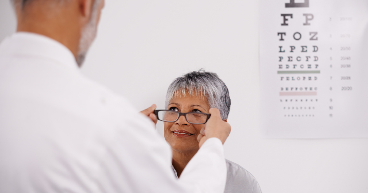
Visual acuity testing measures how clearly someone can see at specific distances. It is an important part of an exam, but it does not provide a complete assessment of eye health.
A patient may read the chart successfully while still developing a condition that affects the retina, optic nerve, peripheral vision, or internal blood vessels. The Centers for Disease Control and Prevention explains that some eye diseases can remain unnoticed during their early stages. Comprehensive examinations help eye doctors identify concerning changes when intervention may be more effective.
Depending on the patient’s age, symptoms, medical history, and risk factors, an exam may include:
Not every patient requires the same combination of tests. Technology is most valuable when it is selected according to a specific clinical need.
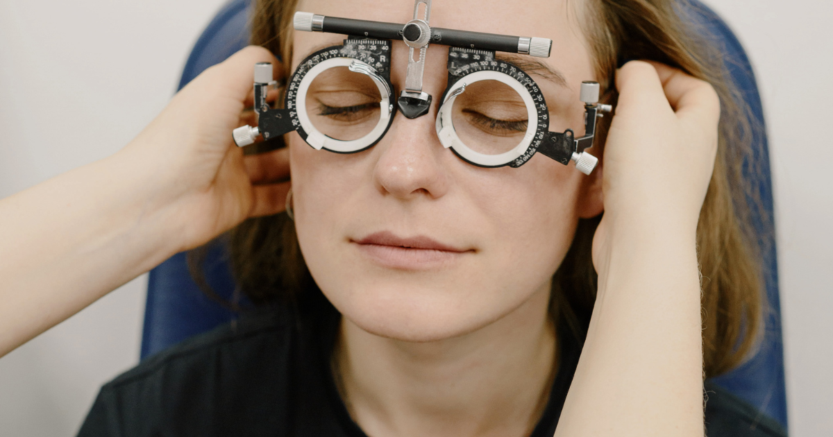
Refraction is the process used to determine whether lenses can improve a patient’s vision. During this part of the exam, the patient compares different lens choices while the eye doctor refines the optical power needed for clearer sight.
The resulting prescription may account for nearsightedness, farsightedness, astigmatism, or presbyopia. The National Eye Institute describes refractive errors as problems that occur when the shape of the eye prevents light from focusing properly on the retina.
Automated instruments can collect useful preliminary measurements, but a prescription is not generated from one number alone. The eye doctor must consider the patient’s responses, visual demands, previous prescription, comfort, and ability to use both eyes together.
This distinction is important because the strongest prescription is not always the most comfortable or practical. A person who spends most of the day working across several digital screens may have different needs from someone who drives frequently, reads fine print, or regularly shifts between near and distant tasks.
Retinal photography captures images of structures at the back of the eye. These images may show the retina, optic nerve, macula, and visible blood vessels. They provide documentation that can be reviewed during the appointment and, when appropriate, compared with future images.
This ability to compare information over time is one of the most useful advantages of digital eye care. A single image represents one moment. A sequence of images may reveal whether an area appears stable or has changed.
Photographs do not independently diagnose every condition, and imaging does not eliminate the need for a complete examination. Instead, it adds another layer of information that the eye doctor can interpret alongside symptoms, medical history, vision testing, and direct observation.
The value comes from integration. Technology captures the image, while the clinician determines what the image means for the individual patient.
During a dilated eye exam, drops temporarily widen the pupils so the doctor can examine more of the eye’s internal structures. The National Eye Institute’s guide to dilated examinations explains that dilation helps doctors check for conditions such as diabetic retinopathy, glaucoma, and age-related macular degeneration.
Dilation may temporarily cause light sensitivity and blurry near vision. Patients may want to bring sunglasses and ask their eye doctor whether they should arrange transportation afterward.
How often dilation is recommended depends on individual risk. Age, diabetes, high blood pressure, family history, previous findings, and certain symptoms may influence the schedule. A personalized recommendation is more useful than assuming everyone needs exactly the same examination interval.
Tonometry measures pressure inside the eye. This measurement can provide important information during a glaucoma evaluation, but one pressure reading does not tell the entire story.
The National Eye Institute notes that some people with elevated eye pressure never develop glaucoma, while others can experience glaucoma-related damage despite having pressure measurements within a generally expected range.
That is why eye pressure may be considered alongside the appearance of the optic nerve, peripheral vision test results, corneal characteristics, family history, and other clinical findings. Technology supplies measurements, but diagnosis depends on how those measurements relate to one another.
The eye offers a direct view of blood vessels and nerve tissue, giving eye doctors an opportunity to observe changes that may be associated with broader health conditions. Diabetes and high blood pressure are especially relevant because they can affect blood vessels within the retina.
A comprehensive eye exam is not a substitute for primary medical care, laboratory testing, or treatment from another healthcare provider. However, an unexpected ocular finding may prompt further evaluation or communication with the patient’s medical team.
For people with diabetes, regular examinations are particularly important. The American Diabetes Association explains that diabetes-related eye disease may be present before a person notices changes in vision. Dilated examination and retinal imaging can help detect signs that require monitoring or treatment.
This connection between eye health and overall health makes an accurate medical history essential. Patients should tell their eye doctor about diagnosed conditions, medications, recent health changes, and relevant family history.
A measurement becomes more informative when it can be compared with earlier results. Digital records allow eye care teams to track prescription changes, eye pressure readings, visual field results, imaging, symptoms, and clinical observations across multiple visits.
Small differences do not always indicate disease. Equipment, testing conditions, patient responses, and normal biological variation can influence results. The clinician’s job is to determine whether a change is expected, requires confirmation, or deserves closer attention.
This is one reason consistency matters. Returning for recommended examinations creates a more complete history, giving the eye doctor additional context for interpreting current findings.
Geek Practitioners has explored this broader shift in its discussion of how technology is advancing modern eye exams. The central benefit is not simply collecting more data. It is using relevant data to support earlier recognition, clearer patient education, and more informed decisions.
Children, working-age adults, contact lens wearers, and older adults do not necessarily have identical visual needs.
For children, an examination may consider visual clarity, focusing, eye alignment, and concerns that could affect learning or development. Adults may notice eye strain, headaches, blurred vision, or difficulty shifting focus after long periods of near work. Later in life, monitoring may place greater emphasis on cataracts, glaucoma, macular changes, and other age-related concerns.
Contact lens wearers generally need additional evaluation because contact lenses interact directly with the eye’s surface. The fit, movement, comfort, prescription, wearing schedule, and corneal response may all need to be assessed.
The American Optometric Association recommends comprehensive care based on a person’s age, health, symptoms, and individual risk factors rather than relying on vision screenings alone.
Patients can help their eye doctor interpret test results by arriving with accurate information. Useful items include current glasses, contact lens details, a medication list, family eye-health history, and information about previous surgeries or treatment.
It also helps to describe symptoms precisely. Instead of reporting that vision is “not right,” consider whether the problem involves distance, reading, glare, double vision, dryness, fluctuating clarity, headaches, flashes, or floaters. Note when the symptoms began and whether they affect one eye or both.
Questions are valuable too. Patients may want to ask:
Routine appointments support preventive care, but sudden symptoms may require prompt attention. Eye pain, injury, abrupt vision loss, new flashes, a sudden increase in floaters, double vision, severe redness, or a curtain-like shadow should not be postponed until the next scheduled examination.
Clarity Eyecare provides comprehensive eye examinations from its office in Baltimore’s Mount Washington neighborhood. Its approach includes carefully measured prescriptions, retinal photography, diagnostic equipment, and evaluation for common eye diseases and health-related changes.
The most important innovation in eye care is not one instrument or image. It is the ability to combine technology, clinical knowledge, patient history, and ongoing comparison. When those elements work together, an eye exam becomes more than a prescription check—it becomes a structured assessment of vision today and a useful reference point for protecting it tomorrow.