Diabetic Eye Exam in Ashburn, VA: Technology for Earlier Detection
A diabetic eye exam in Ashburn, VA can help detect retinal changes before they begin interfering with a patient’s vision. Modern diabetic eye care combines…

A diabetic eye exam in Ashburn, VA can help detect retinal changes before they begin interfering with a patient’s vision. Modern diabetic eye care combines a comprehensive examination with retinal imaging, structured documentation, and coordination between eye doctors and the professionals managing the patient’s diabetes.
This technology-supported approach is important because diabetic eye disease can progress without producing obvious warning signs. Clear vision today does not necessarily mean that the retina is free from early damage.
Diabetes affects the body’s ability to regulate blood glucose. Over time, elevated blood sugar can damage small blood vessels throughout the body, including those supplying the retina—the light-sensitive tissue lining the back of the eye.
Diabetic retinopathy develops when retinal blood vessels weaken, leak, close, or grow abnormally. The National Eye Institute’s diabetic retinopathy overview explains that the condition may eventually cause blurred vision, floating spots, vision loss, or blindness. However, its early stages frequently produce no symptoms.
Diabetes is also associated with several other eye conditions:
A diabetic eye exam evaluates more than whether a patient needs new glasses. Its central purpose is to assess eye health and identify changes requiring monitoring, medical coordination, or treatment.
Many people assume that an eye problem will cause pain, redness, or blurred vision. Early diabetic retinopathy may cause none of these. Small areas of leakage or vascular damage can develop while a person continues to read, drive, and work normally.
The current American Diabetes Association Standards of Care provide evidence-based recommendations for initial retinal examinations and ongoing screening. The appropriate schedule depends on factors such as diabetes type, duration, previous findings, blood glucose management, pregnancy, and the progression of existing retinopathy.
Regular examination creates a sequence of clinical observations. Instead of evaluating a single image or moment, the eye doctor can compare current findings with previous records and determine whether the retina appears stable or has changed.
The exact examination is personalized, but it commonly begins with a review of the patient’s medical and visual history. Patients should be prepared to discuss:
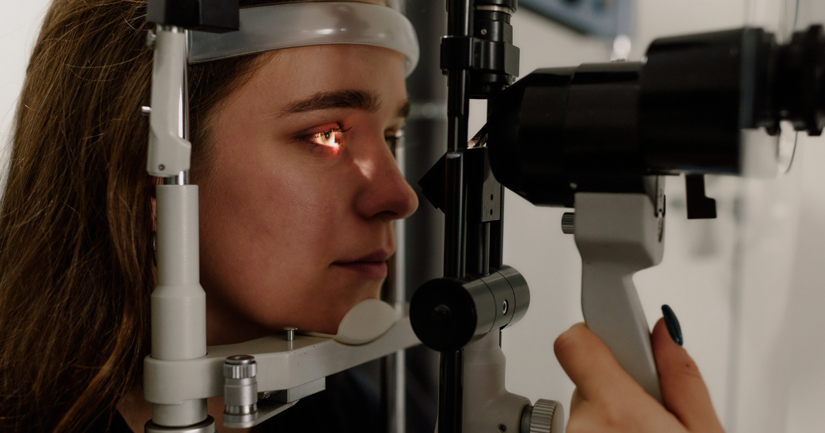
The clinical portion may include visual acuity testing, refraction, evaluation of the pupils, eye-pressure measurement, examination of the front of the eye, and a detailed assessment of the retina and optic nerve.
Dilating drops temporarily enlarge the pupils so the eye doctor can examine a broader portion of the retina. This helps reveal vascular changes, swelling, bleeding, abnormal blood vessels, and other findings that might be difficult to see through an undilated pupil.
The National Eye Institute’s guide to dilated eye examinations explains that dilation supports early detection of diabetic retinopathy, glaucoma, and other conditions that may not initially cause symptoms.
After dilation, vision may remain blurry at near distances, and the eyes may be more sensitive to light for several hours. Patients may want to bring sunglasses and ask whether they should arrange transportation, particularly if they have never been dilated before.
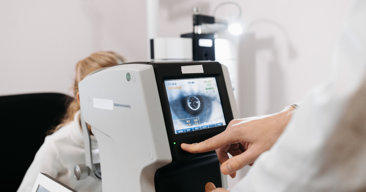
Retinal cameras capture detailed photographs of the structures inside the eye. These images can document the appearance of the optic nerve, macula, blood vessels, and surrounding retina.
Digital photographs provide several practical advantages:
Photography does not automatically replace dilation or a complete examination. Each method supplies different information, and the eye doctor determines which combination is appropriate.
Optical coherence tomography, commonly called OCT, uses reflected light to create high-resolution cross-sectional images of the retina. The technology allows clinicians to examine individual retinal layers and measure their thickness without touching the eye.
OCT can be particularly useful when diabetic macular edema is suspected. It may reveal fluid or structural changes that are not yet obvious to the patient. Repeated scans can also help specialists monitor whether swelling is stable, improving, or progressing.
Not every patient requires OCT at every examination. It is generally ordered when symptoms, retinal findings, or an established diagnosis make the additional information clinically useful.
Artificial intelligence is increasingly used to analyze retinal photographs for signs of diabetic retinopathy. Certain authorized systems can evaluate images and provide a screening result without requiring a specialist to interpret the image initially.
The U.S. Food and Drug Administration has described its authorization of an AI-based device for detecting certain diabetes-related retinal changes. This development demonstrated how automated analysis may expand screening in settings where eye specialists are not immediately available.
AI screening and comprehensive eye care are not identical. An automated result does not evaluate every eye condition, update an eyeglass prescription, measure eye pressure, investigate symptoms, or replace clinical judgment. If screening identifies a concern—or if the image cannot be analyzed—the patient still needs appropriate professional evaluation.
The eyes can provide visible evidence of how diabetes is affecting small blood vessels. For this reason, findings from a diabetic eye exam may be relevant to the patient’s primary care physician, endocrinologist, or diabetes care team.
Secure digital records and structured reports make it easier to share information such as:
The Centers for Disease Control and Prevention emphasizes that optometrists and ophthalmologists play an important role in diabetes care by detecting retinopathy, glaucoma, cataracts, and related conditions.
Coordination does not mean the eye doctor manages every aspect of diabetes. Instead, the examination contributes specialized information to a broader care plan.
Eye examinations are essential, but screening alone cannot prevent every complication. Long-term management of blood glucose, blood pressure, and cholesterol also affects the risk and progression of diabetic eye disease.
The National Institute of Diabetes and Digestive and Kidney Diseases recommends managing the diabetes ABCs:
Personal targets should be established with the healthcare professional managing the patient’s diabetes. Patients should not change medications or treatment goals solely because of general information found online.
Routine examinations are designed to find problems before symptoms appear, but certain changes should not wait until the next scheduled visit. Patients should promptly contact an eye-care professional if they experience:
The American Academy of Ophthalmology provides additional information about diabetic retinopathy symptoms, disease progression, and available treatments. Sudden vision changes may have causes unrelated to diabetes, but they still deserve timely evaluation.
Good preparation helps the eye doctor interpret findings within the patient’s wider health history. Before the appointment:
Patients should also mention previous retinal injections, laser procedures, eye surgery, pregnancy, kidney disease, or major recent changes in blood glucose management.
Ashburn Vision Source provides diabetic eye examinations and ongoing eye care at 44075 Pipeline Plaza, Suite 205, in Ashburn, Virginia. The practice evaluates patients for diabetic retinopathy as well as other conditions associated with diabetes, including cataracts and glaucoma.
The timing of follow-up care depends on the patient’s diabetes history, current eye health, and examination findings. Some patients may remain on a routine screening schedule, while others may require closer monitoring or referral to an ophthalmologist or retinal specialist.
Retinal cameras, OCT systems, digital records, and artificial intelligence are expanding the information available during diabetic eye screening. Their real value, however, comes from using that information within a consistent clinical process.
A careful history provides context. Dilation and examination reveal visible changes. Imaging creates measurable records. Communication connects eye findings to the patient’s broader diabetes care. Together, these elements make it possible to detect problems earlier and respond before avoidable vision loss occurs.