Why the Most Useful Eye Exam Is an Adaptive Workflow
Why the Most Useful Eye Exam Is an Adaptive Workflow Modern personalized eye examinations work like adaptive systems: each answer and finding helps determine what…

Modern personalized eye examinations work like adaptive systems: each answer and finding helps determine what the clinician should investigate next.
That may sound like a technology concept, but it is also a practical way to understand modern eye care. A dependable exam has standardized quality controls, yet it should not feel like the same checklist delivered to every person. The visual demands of an accountant, delivery driver, student, machinist, and recreational athlete are different. So are the risks associated with diabetes, contact lens wear, family history, medication use, and previous eye findings.
The goal is not to perform the greatest possible number of tests. It is to collect the right information, interpret the results together, and produce a clear decision.
In well-designed systems, standardization protects consistency while personalization improves relevance. Eye care follows the same principle. Core elements create a reliable baseline, and the clinician expands or redirects the evaluation when the history, symptoms, or initial measurements justify it.
The American Optometric Association’s comprehensive adult examination guideline presents the exam as a combination of patient history, visual function, ocular health assessment, diagnosis, education, and management. That is much broader than reading an eye chart.
A personalized process might preserve the same clinical foundation while changing:
“My vision is blurry” is useful, but the workflow becomes more precise when that statement is unpacked. Is the blur constant or intermittent? Does it affect one eye or both? Is it worse at a computer, while reading, across a room, or when driving after dark? Did it begin suddenly or gradually? Does blinking change it?
These details help separate problems that may feel similar. A prescription change, an unstable tear film, eye coordination difficulty, cataract, medication effect, and retinal condition can all influence visual quality, but they do not call for the same response.
Bring concrete examples of what is difficult. “Street signs become unclear before I can react” gives different information from “small spreadsheet text loses focus after two hours.” For screen-heavy work, note the monitor distance, number of displays, font size, lighting, and whether symptoms build through the day.
The American Optometric Association’s overview of computer vision syndrome explains that viewing distance, posture, lighting, glare, and uncorrected vision problems can all contribute to digital eye strain. The solution may therefore involve more than changing lens power.
Two people with equally clear distance vision may need different evaluations. A family history of glaucoma, diabetes, high blood pressure, previous eye trauma, long-term medication use, or a new shower of floaters changes the clinical question.
Patients can improve data quality by bringing:
Diabetes illustrates why this information matters. The Centers for Disease Control and Prevention states that people with diabetes have higher risks of retinopathy, glaucoma, cataracts, and vision loss, and it emphasizes comprehensive exams that include dilation. A routine vision concern can therefore become part of a broader health-monitoring pathway.
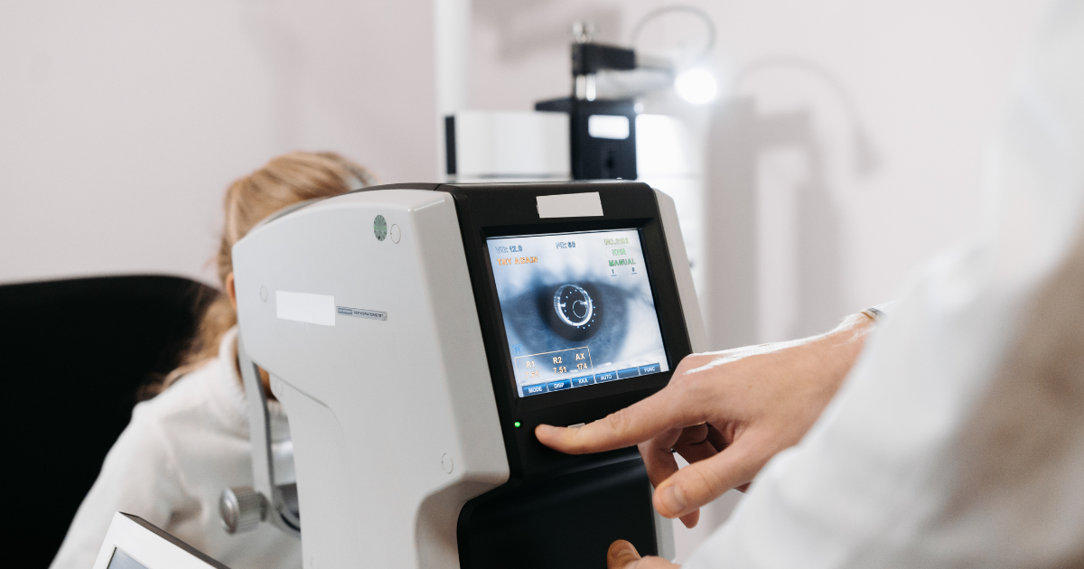
Digital instruments can capture precise numbers and detailed images, but every output still needs context. Visual acuity records clarity under defined conditions. Refraction estimates the lens power that produces the best response. Tonometry measures pressure inside the eye. Imaging records visible structures. Each contributes a piece of evidence rather than a complete conclusion.
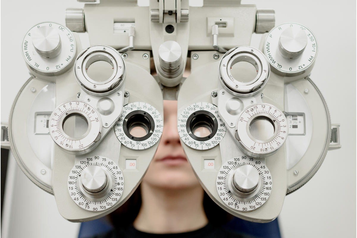
The familiar “Which is better, one or two?” sequence is not a test of whether the patient can give perfect answers. It is an iterative comparison. Small differences may genuinely look the same, and saying so is useful. The final prescription should also be considered alongside comfort, binocular balance, working distance, previous correction, and adaptation needs.
The National Eye Institute’s guide to refractive errors explains that nearsightedness, farsightedness, astigmatism, and presbyopia arise when the eye does not focus light correctly on the retina. The same “blur” label can therefore represent several optical patterns.
A pressure reading is important, but it should not be interpreted alone. The National Eye Institute notes that some people with elevated pressure never develop glaucoma, while some people with pressure in the statistically normal range do. Optic nerve appearance, visual field results, risk factors, and change over time may all affect the assessment.
This is a good example of why personalization is not a luxury feature. The meaning of the number depends partly on the person to whom it belongs.
Retinal imaging can create a detailed visual record that supports documentation, patient education, and comparison over time. It can also reveal findings that warrant a closer look. Its value grows when the clinician connects the image with symptoms, examination findings, and prior records.
Patients should not assume that every camera image is interchangeable with pupil dilation. The National Eye Institute’s explanation of dilated exams says dilation lets the doctor examine internal eye structures and notes that additional tests may be included according to individual needs. The clinician should decide when imaging, dilation, or both are appropriate.
A useful question is not simply, “Did the office use advanced technology?” Ask, “What did this test evaluate, what did it show, and will today’s result become a baseline for future comparison?”
One exam shows the current state. A series of comparable exams can reveal direction and rate of change. A small shift in prescription may be unremarkable in one context and meaningful in another. The same principle applies to eye pressure, optic nerve appearance, retinal findings, visual fields, and symptoms.
Continuity depends on accurate records and consistent comparisons. Tell the clinician if a test was performed elsewhere, if a medication recently changed, or if a symptom resolved and returned. When possible, bring prior prescriptions or request that relevant records be transferred before the visit.
Even excellent testing loses value if the patient leaves without understanding the result. A practical explanation should distinguish among a normal finding, a monitored variation, a working diagnosis, and an urgent concern. It should also clarify what happens next and which symptoms should prompt earlier contact.
The Agency for Healthcare Research and Quality’s teach-back guidance recommends asking patients to explain important information in their own words. This is not a quiz. It is a way to confirm that instructions were communicated clearly.
Before leaving, a patient should be able to answer:
Fort Worth Eye Center describes its complete exams as assessments of prescription, ocular health from front to back, binocular vision, and minor neurological function. Its published exam process also incorporates iCare EIDON widefield TrueColor confocal fundus imaging as a required screening component.
That combination illustrates the central idea: technology is most useful when it contributes to a connected evaluation rather than standing alone. A retinal image, refraction result, pressure measurement, and symptom history become more informative when one clinician interprets how they relate.
The practice states that exam frequency may vary according to age, medical history, and the use of glasses or contact lenses. That risk-based approach is consistent with the CDC’s vision-health guidance, which recommends discussing individual timing when conditions such as diabetes or high blood pressure are present.
A modern eye exam is not valuable merely because it produces more data. Its value lies in converting selected data into a decision the patient can use.
That output may include a prescription, recommendations for a specific visual task, monitoring of a baseline finding, treatment, or referral. Federal consumer protections also support portability: the Federal Trade Commission’s Eyeglass Rule requires prescribers to provide a copy of an eyeglass prescription after a refractive exam without an extra charge.
The best exam workflow is therefore both technically sound and personally relevant. It begins with the right questions, adapts to evidence, explains uncertainty, and ends with clear next steps. That is what turns a sequence of tests into meaningful eye care.