Why a Modern Eye Exam Is More Than a Prescription Check
Comprehensive eye examinations combine vision testing, health assessment, diagnostic technology, and clinical interpretation to create a more complete picture of how the eyes are functioning.
Many people visit an eye doctor because road signs have become harder to read or computer text seems less distinct. Updating a prescription is important, but it represents only one part of a thorough examination. The larger purpose is to evaluate vision quality, inspect eye structures, identify risk factors, and establish measurements that can be compared in the future.
This makes an eye exam similar to preventive maintenance in a well-managed organization. Waiting until a system fails is usually more disruptive than identifying a developing issue early. The same principle applies to vision, particularly because several significant eye diseases can progress without obvious warning signs.
Clear Vision Does Not Always Mean Healthy Eyes
A person can read the smallest line on an eye chart and still have an emerging eye-health concern. Visual acuity measures how clearly someone sees at a particular distance, but it does not independently evaluate the retina, optic nerve, eye pressure, peripheral vision, or other structures.
The National Eye Institute explains that many eye diseases produce few or no early symptoms. A dilated examination allows an eye doctor to see more of the inner eye and look for changes before the patient notices vision loss.
This distinction is important when comparing a basic vision screening with a comprehensive exam. A screening may flag reduced visual acuity or another potential issue, but it is not designed to provide the same depth of evaluation. The American Academy of Ophthalmology’s eye-exam overview describes how a complete evaluation can include multiple tests selected according to age, symptoms, medical history, and risk.
What the Different Tests Actually Measure
A modern eye examination is not one test. It is a collection of measurements and observations that answer different questions. Findings are interpreted together because one result rarely tells the entire story.
Visual Acuity
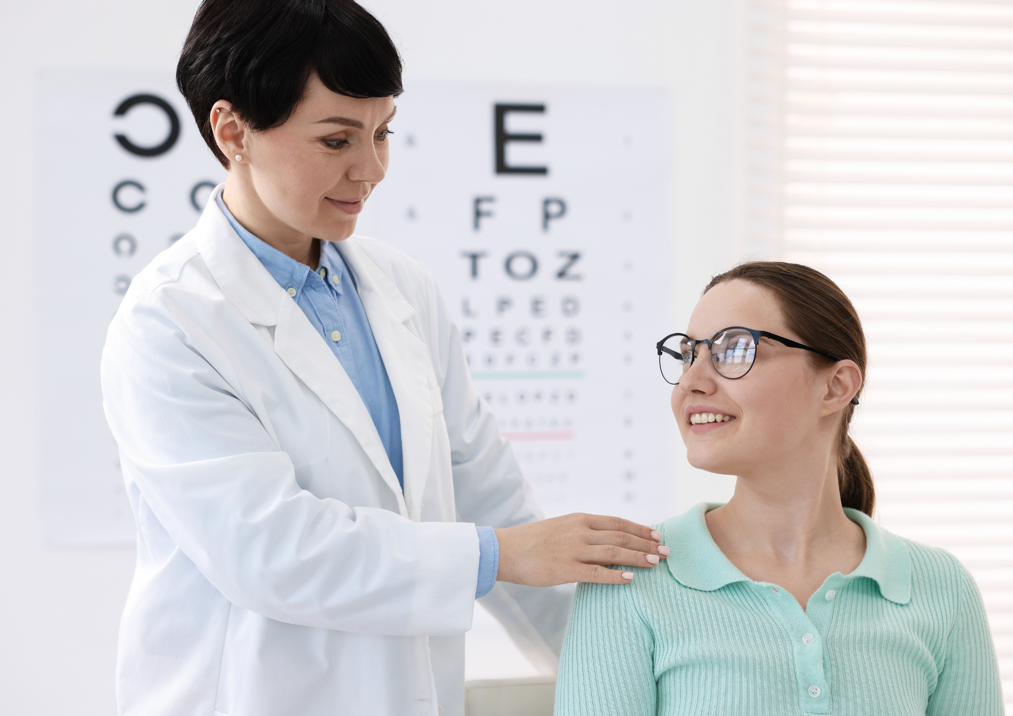
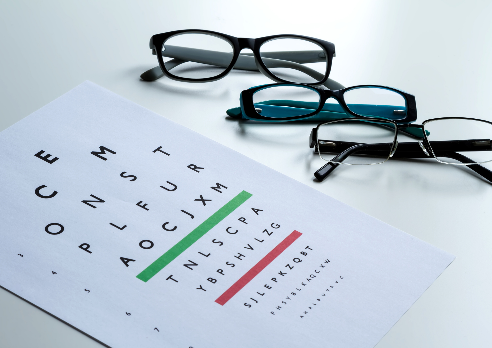
The familiar letter chart measures how clearly each eye sees at specified distances. Testing the eyes individually can reveal differences that may be less noticeable when both eyes are open.
Refraction and Retinoscopy
Retinoscopy provides an objective estimate of refractive error by observing how light reflects from the eye. Refraction then helps refine the lens power needed for glasses. During the familiar “one or two” portion of the exam, the patient compares lens options so the doctor can determine which produces clearer, more comfortable vision.
Refraction can identify nearsightedness, farsightedness, astigmatism, and presbyopia. The goal is not merely to produce the sharpest possible line of letters. A useful prescription should also reflect the patient’s working distance, visual demands, and ability to use both eyes comfortably.
Visual Field Testing
A visual field assessment evaluates peripheral vision. Depending on the method used, the patient may respond when lights or targets appear away from the center of view. Blind spots or unusual patterns may require repeat testing or further investigation.
Tonometry
Tonometry measures pressure inside the eye. Elevated pressure can increase glaucoma risk, but pressure alone does not confirm or exclude the disease. Some people with higher pressure never develop optic nerve damage, while others develop glaucoma at statistically normal pressure levels.
The National Eye Institute’s glaucoma resource emphasizes the value of combining pressure measurement with optic nerve evaluation and visual field testing.
Slit-Lamp Examination
A slit lamp provides a magnified view of structures at the front of the eye. It helps the doctor examine areas such as the cornea, iris, lens, eyelids, and tear film. This portion of the exam may reveal signs of cataracts, corneal conditions, inflammation, or dry eye.
Retinal and Optic Nerve Evaluation
Ophthalmoscopy, dilation, and retinal imaging can provide information about the retina, optic nerve, macula, and visible blood vessels. Digital images are especially valuable as documentation. Future images can be compared with earlier records to determine whether an area appears stable or has changed.
Technology Improves Visibility, Not Clinical Judgment
Modern diagnostic equipment can capture details that are difficult to document through description alone. High-resolution retinal images, pressure measurements, visual field data, and other test results create a structured clinical record.
Technology is most useful when it supports interpretation rather than replacing it. An image may identify an unusual area, but an eye doctor must consider whether it represents normal variation, an imaging artifact, or a finding that deserves additional evaluation.
The strongest decisions come from combining several information sources:
- Current test results
- Previous images and measurements
- Changes reported by the patient
- Family eye-health history
- Medications and medical conditions
- Work, school, driving, and recreational demands
- Clinical examination of the eyes
This layered approach reduces dependence on any single number or image. It also creates a better baseline for long-term care.
The Eyes Can Offer Clues About General Health
The eye is unusual because its transparent structures allow clinicians to directly observe blood vessels and nerve tissue. An eye exam does not replace care from a primary physician, but certain findings may suggest that additional medical evaluation is appropriate.
The Centers for Disease Control and Prevention notes that eye examinations may reveal signs associated with conditions such as diabetes and high blood pressure. These observations should be communicated to the patient and, when appropriate, coordinated with another healthcare provider.
Diabetes is particularly relevant because prolonged high blood sugar can damage retinal blood vessels. The National Eye Institute’s diabetic retinopathy guidance explains that early stages may not cause symptoms, making recommended retinal examinations important even when vision seems unchanged.
Why Historical Data Matters
One examination is a snapshot. Several examinations create a timeline.
A mild variation may not be concerning by itself, but a consistent pattern of change can be clinically meaningful. Comparing prescriptions, pressure readings, retinal images, visual fields, and optic nerve appearance helps the doctor determine whether the eyes remain stable.
This is why keeping regular records with an eye-care provider can be valuable. The purpose is not to repeat the same process without reason. It is to create comparable information that makes subtle trends easier to recognize.
At Luminary Eye Care in Marietta, Georgia, comprehensive exams are personalized according to medical history, visual needs, and lifestyle. The practice uses diagnostic technology to evaluate visual acuity, prescription needs, eye pressure, peripheral vision, retinal health, and internal eye structures. This broader approach supports both clear vision and preventive eye care for patients in Marietta and East Cobb.
Who May Need More Frequent Eye Examinations?
There is no single examination schedule that is appropriate for everyone. Frequency depends on age, symptoms, family history, health conditions, medications, previous findings, and whether the person wears corrective lenses.
The American Optometric Association encourages regular comprehensive eye care because vision and eye health can change throughout life.
Closer monitoring may be recommended for people who:
- Have diabetes or high blood pressure
- Have a family history of glaucoma or macular degeneration
- Use medications that may affect the eyes
- Wear contact lenses
- Have experienced an eye injury or surgery
- Have high or changing prescriptions
- Have previously identified retinal, corneal, or optic nerve findings
- Notice new difficulty reading, driving, or working at a screen
Age also changes risk. Children need evaluation as their visual system develops, while adults may experience presbyopia and changing occupational demands. Later in life, the likelihood of cataracts, glaucoma, and age-related macular degeneration increases. The National Eye Institute provides additional information about how macular degeneration can affect central vision.
How to Prepare for a More Productive Exam
Accurate information helps the doctor interpret test results in context. Before the visit, gather details that may affect vision or eye health.
- Bring current glasses and contact lenses.
- List prescription medicines, over-the-counter products, and supplements.
- Note any family history of glaucoma, retinal disease, or significant vision loss.
- Describe symptoms precisely, including when they started and what makes them better or worse.
- Mention the amount of time spent driving, reading, using digital devices, or performing detailed work.
- Ask whether dilation is expected and bring sunglasses if recommended.
- Arrange transportation when dilation or another procedure may temporarily affect vision.
It is also helpful to describe visual challenges rather than simply saying a prescription feels wrong. Difficulty with night driving, fluctuating clarity, headaches after close work, glare, dryness, or trouble shifting focus between distances can provide useful clues.
When Not to Wait for a Routine Exam
Regular care is preventive, but certain symptoms warrant prompt attention. Sudden vision loss, new flashes or a sudden increase in floaters, significant eye pain, a curtain-like shadow, chemical exposure, trauma, pronounced redness, or new double vision should be evaluated urgently.
The CDC’s eye-health recommendations advise contacting an eye-care professional for symptoms such as decreased vision, eye pain, drainage, double vision, flashes, floaters, or halos around lights.
A Better Way to Think About Routine Eye Care
A comprehensive exam is not simply a transaction that ends when a glasses prescription is printed. It is a structured review of vision performance, ocular health, personal risk, and change over time.
Diagnostic technology makes more information available, but its value depends on careful interpretation and communication. Patients contribute by sharing accurate histories, describing how they use their eyes, and returning at the interval recommended for their individual needs.
When those elements work together, an eye examination becomes a practical form of preventive maintenance. It can improve day-to-day visual performance while building the records needed to protect sight over the long term.